


Osteoarthritis of the hip joint is a degenerative-dystrophic pathology characterized by destruction of hyaline cartilage.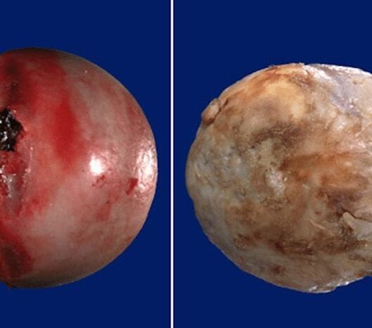 The disease develops gradually, accompanied by pain and decreased mobility. In the absence of medical intervention in the early stages of osteoarthritis, atrophy of the thigh muscles occurs after a few years.
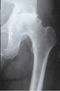 The injured limb shortens, and the fusion of the joint space causes partial or complete immobilization of the hip joint. The causes of pathology are previous injuries, curvature of the spine, systemic diseases of the musculoskeletal system.
Osteoarthritis is commonly found in middle-aged patients and the elderly. Diagnosis is based on the results of instrumental studies - radiography, MRI, CT, arthroscopy. Treatment of grade 1 and 2 severity pathology is conservative. If ankylosis is detected or drug treatment is ineffective, surgery (arthrodesis, endoprosthesis) is performed.
The mechanism of development of pathology
The hip joint is made up of two bones - the ilium and the femur. The lower part of the ilium is represented by the body, which is involved in the articulation of the thigh, forming the upper part of the acetabulum. During movement, the glenoid fossa is immobile and the femoral head moves freely. Such a "hinge" device of the hip joint allows you to bend, bend, rotate, encourages the hip to be flexed and attached. The smooth, durable, long-lasting hyaline that straightens the acetabulum and femoral head ensures unimpeded sliding of cartilage and joint structures. Its main functions are redistribution of loads during movement, preventing rapid wear of bone tissue.
Cartilage trophism is impaired under the influence of external or internal factors. It does not have its own circulatory system - it provides nutrients to the synovial fluid tissue. It thickens with osteoarthritis and becomes sticky.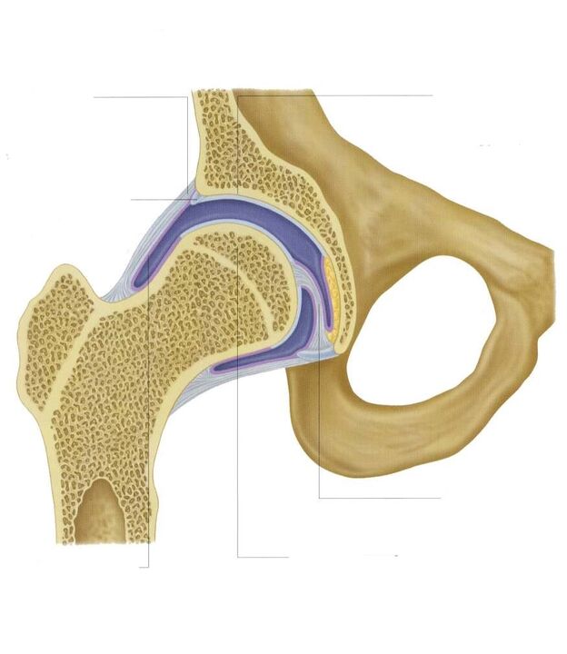 The resulting malnutrition causes the surface of the hyaline cartilage to dry out. It is covered with cracks, which cause permanent microtraumas of the tissues during bending or stretching of the hip joint. Cartilage thins and loses its cushioning properties. The bones are deformed to "adapt" to the increase in pressure. And destructive and degenerative changes are advancing against the background of deterioration of metabolism in tissues.
Causes and motivating factors
Idiopathic or primary osteoarthritis develops for no apparent reason. It is believed that the destruction of cartilage tissue is due to the natural aging of the body, slowing down the recovery process, a decrease in the production of collagen and other compounds necessary for the complete restoration of the structures of the hip joint. Secondary osteoarthritis occurs against the background of an already existing pathological condition in the body. The most common causes of secondary disease are:
- previous injuries - damage to the ligament-tendon apparatus, muscle fractures, complete separation from the bone base, fractures, dislocations;
- joint developmental disorders, congenital dysplastic diseases;
- autoimmune pathologies - rheumatoid, reactive, psoriatic arthritis, systemic lupus erythematosus;
- non-specific inflammatory diseases such as purulent arthritis;
- specific infections - gonorrhea, syphilis, brucellosis, ureaplasmosis, trichomoniasis, tuberculosis, osteomyelitis, encephalitis;
- disorders of the endocrine system;
- degenerative-dystrophic pathologies - osteochondropathy of the femoral head, osteochondritis dissections;
- Hypermobility of the joints leads to the production of "super stretching" collagen, excessive mobility, weakness of the ligaments.
Hematopoietic disorders are among the provoking factors, as the cause of the development of osteoarthritis may be hemarthrosis (bleeding in the cavity of the hip joint). The initial conditions of the disease are overweight, excessive physical activity, sedentary lifestyle. Improper organization of sports training, its development is caused by a deficiency in the diet of foods with high amounts of micronutrients, fat and water-soluble vitamins. Postoperative osteoarthritis occurs several years after surgery, especially if accompanied by the removal of large amounts of tissue. Hyaline cartilage trophism is often associated with hypothermia, living in an environment that is unfavorable to the environment, and working with toxic substances.
Osteoarthritis of the hip joint can not be inherited. However, in the presence of certain congenital features (metabolic diseases, skeletal structure), the likelihood of development increases significantly.
Symptoms
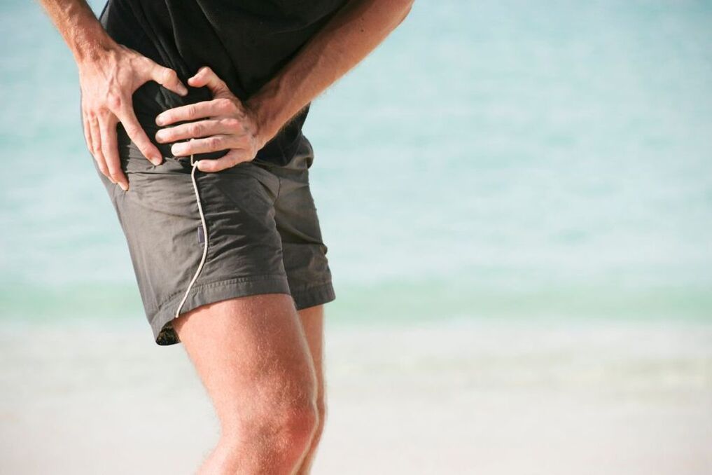
The leading symptoms of osteoarthritis of the hip joint are pain when walking in the thigh area, spread to the groin, knee joint. A person suffers from stiffness, stiffness of movements, especially in the morning. To stabilize the joint, the patient begins to slow down, changing gait. Over time, the limb shrinks significantly due to muscle atrophy and articulation deformity. Another characteristic feature of the pathology is the limitation of thigh abduction. For example, difficulties arise when trying to sit on a separate stool with one's feet.
Periodic pain occurs after intense physical exertion for osteoarthritis of the first severity. They are localized in the joint area and disappear after a long rest.
The severity of the pain syndrome increases with secondary arthrosis of the hip joint. Anxiety occurs even at rest, extends to the thigh and groin area, and increases with weight lifting or increased motor activity. A person begins to feel difficulty in relieving pain in the hip joint. Limitation of movement in the joint is noted, especially during thigh abduction and internal rotation.
Tertiary osteoarthritis is characterized by persistent severe pain that does not decrease day and night. There are difficulties in moving, so a person has to use a stick or crutch while walking. The hip joint is stiff, there is significant atrophy of the thigh, thigh and leg muscles. Due to the weakness of the abductor femoral muscles, the pelvic bones are displaced in the frontal plane. To compensate for the shortening of the leg, the patient bends towards the injured limb while moving. This causes a strong change in the center of gravity and an increase in stress on the joint. At this stage of osteoarthritis, clear ankylosis of the joint develops.
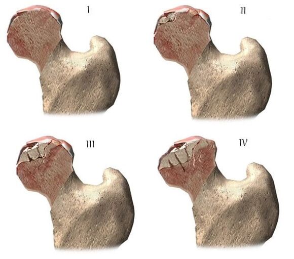
| Degrees | Radiographic signs |
| First | No changes will be announced. The joint cavities narrow moderately, unevenly, and there is no destruction on the surface of the thigh. Small bone growths are observed on the outer or inner edge of the acetabulum |
| The second | Due to the uneven combination, the height of the joint space is significantly reduced. The bony head of the femur moves upwards, deforms, enlarges, and the contours are uneven. Bone growths form on the surface of the inner and outer edges of the glenoid fossa |
| Third | There is a complete or partial combination of the joint space. The femoral head expands strongly. Numerous bone growths are located on all surfaces of the acetabulum |
Diagnostics
When making a diagnosis, the doctor takes into account the pathology, anamnesis, the results of external examination of the patient and the clinical manifestations of instrumental studies. Radiography is the most informative. With its help, the condition of the hip joint is assessed, the stage of development, the degree of damage to cartilage tissue and, in some cases, the cause of development are determined. If the cervico-diffuse node is enlarged and the acetabulum is bent and straightened, it is likely that dysplastic congenital changes in articulation can be assumed. Perthes' disease, or young epiphysiolysis, is indicated by a deformed form of the hip bone. Radiography can detect post-traumatic osteoarthritis, even if there is no previous history of trauma. Other diagnostic methods are also used:
- CT helps to detect the growth of osteophytes on the edges of bone plates;
- MRI is performed to assess the condition of connective tissue structures and the degree of involvement in the pathological process.
If necessary, the inner surface of the joint is examined with arthroscopic instruments. Differential diagnosis is made to exclude gonarthrosis, lumbosacral or thoracic osteochondrosis. Pain in osteoarthritis can be disguised as a clinical manifestation of radicular syndrome caused by seizures or inflammation. With the help of a number of tests it is possible to exclude neurogenic pathology in general. Osteoarthritis of the hip joint is definitely different from trochanteric bursitis of the hip joint, ankylosing spondylitis, reactive arthritis. Biochemical studies of blood and synovial fluid are performed to rule out autoimmune pathologies.
Drug treatment tactics
Medical treatment is aimed at improving the patient's well-being. Drugs of different clinical and pharmacological groups are used for this purpose:
- Non-steroidal anti-inflammatory drugs (NSAIDs) - Nimesulide, Ketoprofen, Diclofenac, Ibuprofen, Meloxicam, Indomethacin, Ketorolac. Injectable solutions are used to relieve acute pain, and pills, pills, ointments, gels help relieve pain of mild to moderate severity;
- Glucocorticosteroids - Triamcinolone, Dexamethasone, Hydrocortisone. Procaine is used in combination with lidocaine anesthetics in the form of intraarticular blockade;
- muscle relaxants - Baclofen, Tizanidine. Skeletal muscle spasms are included in treatment regimens for squeezing sensitive nerve endings;
- Drugs that improve blood circulation in the joints - Nicotinic acid, Aminophylline, Pentoxifylline. It is prescribed to patients to improve tissue trophism and prevent the development of the disease;
- chondroprotectors. It is effective only in the 1st and 2nd stages of osteoarthritis.
Rubbing ointments with a warming effect helps to relieve mild pain. The active ingredients of foreign agents are capsaicin, cinquefoil, camphor, menthol. These substances are characterized by local irritating, distracting, analgesic effects. Compressed joints with dimethylsulfoxide will help to cope with medical bile, edema, morning swelling of the thighs. Patients are recommended classic, acupressure or vacuum massage for coxarthrosis. Daily exercise therapy is an excellent prevention of further osteoarthritis.
Surgical intervention
An operation is performed with a pathological diagnosis of ineffectiveness of conservative treatment or a complication of ankylosis. It is not possible to repair cartilage tissue in a joint damaged by osteoarthritis without prosthetic surgery, but the right approach to treatment, adherence to all medical prescriptions, maintaining a healthy lifestyle, therapeutic exercise, regular massage courses, vitamin intake and proper nutrition, cartilage and hip joint lesion processand you can stop the destruction.